5 Thymus
The thymus is responsible for the training of T-cells, ensuring that self-reactive T-cells are removed from circulation. The thymus is at its largest and most active in neonatal and young animals, where it forms a large, multilobular, intrathoracic organ in the cranial mediastinum. As you’ll recall, the thymus regresses over the lifetime of an animal, becoming practically inapprent by the time most animals have reached sexual maturity. Although still present, at this stage the thymus is typically indistinguishable from the mediastinal fat.
It is broken down into lobules composed of a cortex and medulla, and has both an epithelial and lymphoid component.
With the exception of a few diseases, thymuses tend not to be a primary source of clincial disease.
5.1 Miscelaneous conditions
5.1.1 Atrophy
Thymic atrophy is one of the most common chagnes noted in the thymus. Two basic pathogenic mechanisms can lead to thymic atrophy. Because the lymphoid population of the thymus is compsoed of precursor cells from the bone marrow, destruction of precursor cells in the bone marrow can lead to atrophy of the thymus. The second mechanism is direct damage to the lymphocytes within the thymus itself. Causes include viral infection (e.g. BVDV, canine distemper virus, FIV, canine and feline parvovirus, EHV-1, and classical swine fever virus), toxins, chemotherapy, and radiation. Care must be taken when deciding if the thymus is atrophied: the thymus naturally involutes with age, and thus age-matched controls are often necessary to grossly identify atrophy.
5.1.2 Hypoplasia
Recall that aplasia signifies failure of an organ to reach its normal size (contrast this with atrophy, in which organ has at one point been normal, but has then become smaller). Aplasia of the thymus is a congenital defect, usually involving an immunodeficiency of T cells. The most common manifestation of thymic hypoplasia is part of severe combined immunodeficiency syndrome in foals, particularly Arabians, and certain breeds of dogs (Jack Russell terriers, Bassett hounds). The thymus of these animals is devoid of lymphocytes, rendering them markedly small. These animals are predisposed to infection and frequently do not live long after birth.
5.1.3 Hemorrhage
Massive thymic hemorrhage is an uncommon but important finding particularly in dogs, where it can be the only lesion. Three main causes are implicated, and should be included in a differential list:
- Anticoagulant rodenticides
- Trauma
- Idiopathic
These three causes are indistinguishable on gross examination, and would require additional history and/or toxin testing. The degree of hemorrhage can be profound and life threatening.
5.2 Thymic neoplasia
The thymus has an epithelial and a lymphoid component, and either may give rise to a neoplasm. Neoplastic proliferations of lymphocytes are lymphomas, while in the thymus, epithelial neoplasms are known as thymomas.
5.2.1 Thymic lymphoma
Unsurprisingly, thymic lymphoma is almost always T-cell in origin. It usually affects younger animals, particularly cats, calves, and less commonly dogs. In young cats, thymic lymphoma is highly associated with feline leukemia virus (FeLV), and the widespread vaccination of cats against this virus has dramatically decreased the incidence of the disease in these animals. There is no viral association in other animals, or in older cats.
Grossly, the neoplasm is generally present diffusely throughout the thymus, creating a large, space-occupying mass in the mediastinum. The mass may get large enough that it compresses the lungs, resulting in dyspnea. Thoracic effusion is also frequently present.
5.2.2 Thymoma
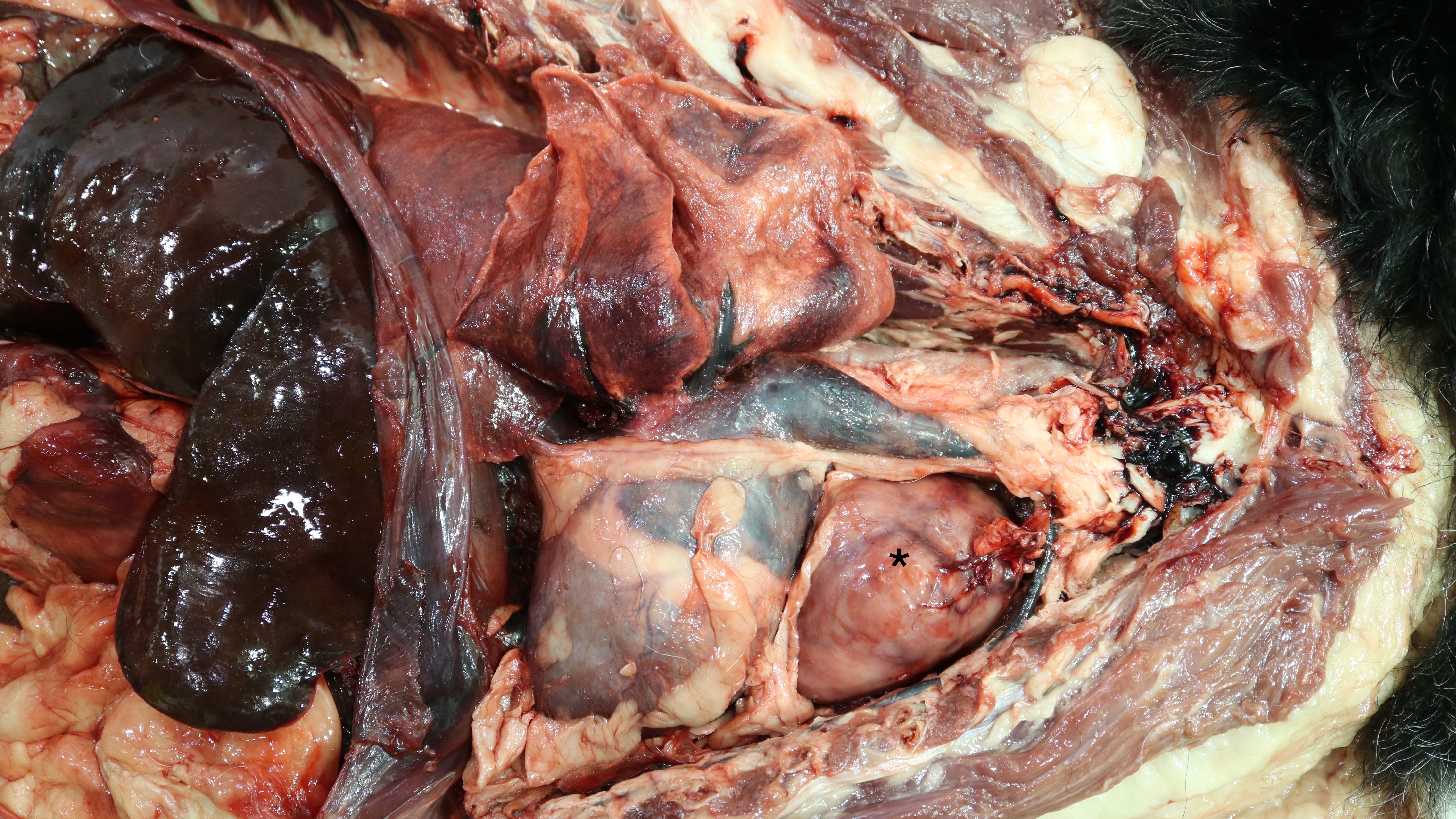
This neoplasm of the epithelial component of the thymus is most frequently seen in dogs and small ruminants (sheep, goats). They tend to be slow-growing, relatively benign tumours that only rarely metastasize. They are grossly indistinguishable from thymic lymphoma, thus, histopathology is required to differentiate between the two (Fig 5.1). Dogs with thymomas often develop myasthenia gravis (note: link takes you to a separate package of course notes on skeletal muscle. The details of myasthenia gravis are not required for exams on the hemolymphatic system).

Figure 5.1: The asterix marks a firm, white, multilobular mass in the cranioventral thorax of this dog. Differential diagnoses include thymoma and thymic lymphoma (histopathology was not performed in this case).