4.1.3.1 Primary neoplasia
Lymphoma, lymphoma, lymphoma. Lymphoma is one of the most common malignant neoplasms of many domestic species. Whether you practice bovine, equine, or companion animal medicine, you will encounter lymphoma. Chickens? Lymphoma. Wildlife? Usually infectious diseases, but sometimes, lymphoma. It is worth taking your time to work through this section (because you will need to know it, and because it will be on the exam). As a sidenote, lymphoma is sometimes referred to as lymphosarcoma. The two terms mean the same thing, and lymphoma is currently the preferred term.
The classification of lymphomas is a complex, multifactorial process, involving clinical, histopathological, immunohistochemical, and molecular information. In reality, the diagnostic process often ends with the diagnosis of ‘lymphoma’, rather than pursuing the advanced diagnostic modalities that would be commonplace in human medicine. The current classification system was developed by the World Health Organization, and includes around 35 different subtypes. Some practical aspects of classification are useful to review.
Anatomical location
- Multicentric: present in multiple lymph nodes, ± liver, spleen, bone marrow
- Cutaneous: in the skin
- Alimentary: within the gastrointestinal tract
- Hepatosplenic: in the liver and/or spleen
- Thymic: Affects the (you guessed it) thymus.
- Miscellaneous: ocular, hepatic, specific splenic forms, etc.
Immunophenotype: B or T cell, or neither (e.g NK cell).
Grade: based on histopathological features, usually mitotic count. A higher grade is associated with a poorer prognosis.
Lymph nodes with lymphoma appear grossly enlarged and homogenously pale white, occasionally streaked with hemorrhage. They are typically firm. It can be difficult to grossly distinguish between reactive hyperplasia and lymphoma (can you think of some features that might help you differentiate between the two?).
There are key species differences when it comes to lymphoma.
4.1.3.1.1 Canine
Lymphoma is the most common hematopoeitic neoplasm of dogs. Large-breed dogs are predisposed, including Golden and Labdrador retrievers and Boxers. Cocker spaniels, Terriers, and Beagles are also overrepresented. Several forms of lymphoma occur in the dog, but multicentric is by far the most common, usually presenting as a generalized lymphadenopathy, especially of the peripheral lymph nodes. They can be either B- (more common) or T-cell in origin. Clinical signs are vague and are often absent during the early stages of disease. Alimentary lymphoma occurs but is less common than in cats. They are usually of T-cell origin, and often progress slowly quickly (edit Sept 14/21). Clinical signs relate to the GI tract: diarrhea, vomiting, and weight loss. Cutaneous lymphomas, most commonly epitheliotropic T-cell lymphoma, are about as common as GI lymphoma. They are often a cause of pruritus, but gross lesions are varied and difficult to distinguish from other causes of pruritus. Epitheliotropic lymphoma may affect the skin or gums (Fig 4.1). The disease is slowly progressive. A variety of other slowly progressive lymphomas affect the dog, but are infrequent. Chemotherapy is available for many canine lymphomas with varying degrees of success, but can sometimes induce remission.

Figure 4.1: A) A poorly demarcated area of the gingival mucosa is deep red to purple, looking very much like an ecchymosis (bruise). B) Histology of this area reveals a neoplastic population of round cells infiltrating the epidermis and superficial dermis. The diagnosis was epitheliotropic lymphoma.
4.1.3.1.2 Feline
Lymphoma is the most common malignant neoplasm of cats, with alimentary lymphoma being the most common anatomical type. Other forms, such as multicentric, nasopharyngeal, or mediastinal, occur, but with far less frequency. Alimentary lymphoma in cats is usually of T-cell origin (formally known as enteropathy-associated T-cell lymphoma). This type of lymphoma typically progresses slowly, with weight loss, diarrhea, and vomitting as the principle clincal signs. The microscopic appearance of the disease is one of infiltrative lymphocytes within the epithelium and lamina propria, and it can therefore be quite difficult to distinguish from inflammatory bowel disease. Submitting full-thickness or deep-endoscopic biopsies gives you the best chance at a diagnosis. Many endoscopic biopsies are ‘wimpy’ - consisting of shallow portions of the superficial mucosa, giving the pathologist little ability to distinguish between the two diseases. The prognosis for this type of lymphoma is a bit controversial, as large scale studies are still unavailable, but it is generally considered to be a slowly ‘smoldering’ disease that an animal may live with for a significant amount of time.
Multicentric lymphoma is the next most common form in the cat, and unlike in the dog, involvement of the peripheral lymph nodes is not a prominant feature. Instead, liver and kidney are more commonly involved.
Historically, feline lymphoma was highly associated with the prevalance of feline leukemia virus, and testing and control of FeLV has greatly reduced the incidence of lymphoma, particularly thymic and multicentric lymphomas, in younger cats. FIV has also been associated with lymphoma.
4.1.3.1.3 Bovine
Lymphoma in cows is separated into two major forms:
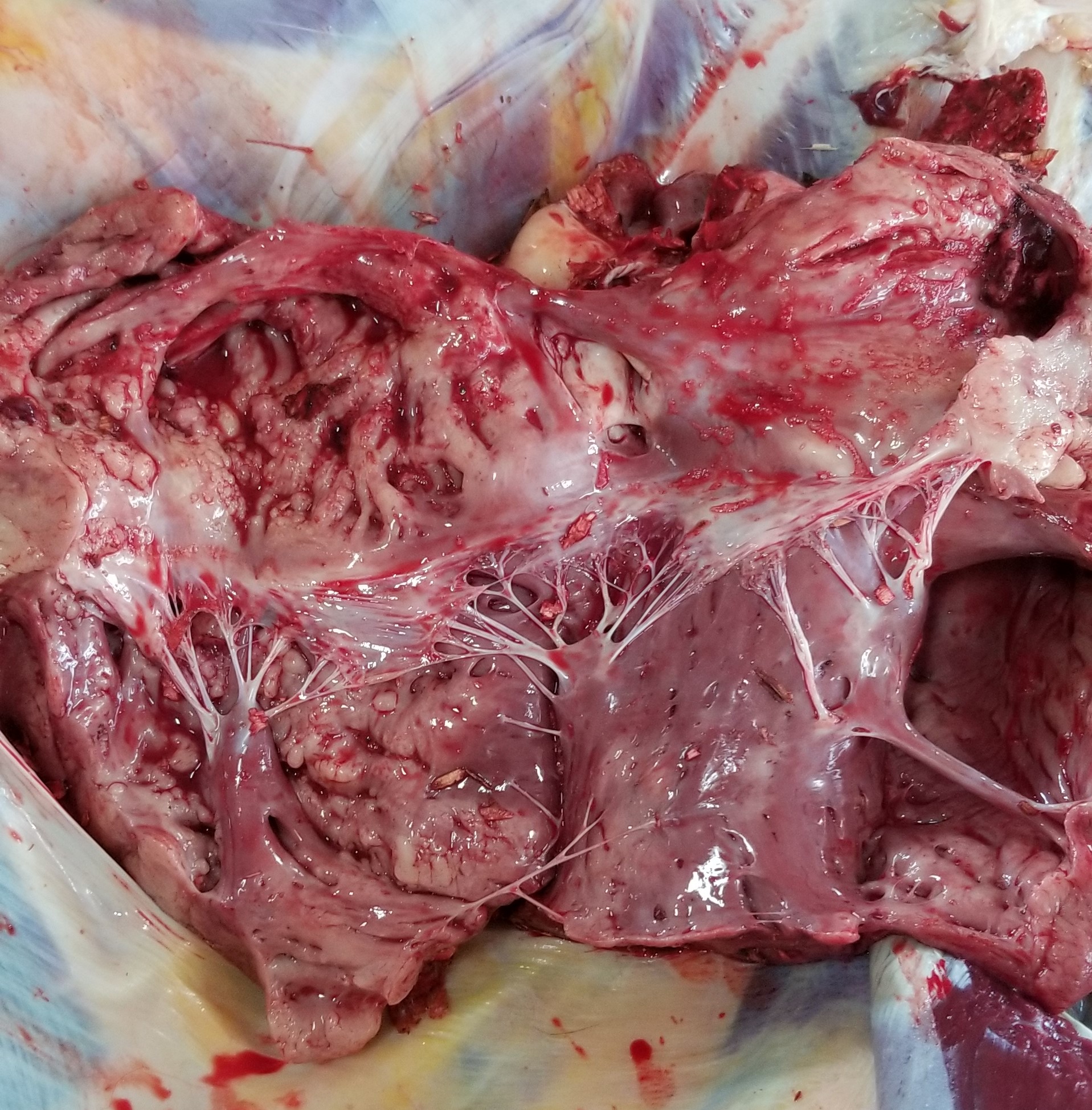
Enzootic bovine leukosis: Caused by bovine leukemia virus (BLV), a retrovirus that infects B-cells. The virus is transmitted horizontally, mostly by infected arthropods, iatrogenically (re-use of needles, rectal sleeves), colostrum/milk administration, or natural breeding. The virus causes multicentric lymphoma in cattle 5-8 years old. Like all retroviruses, once the virus has infected the host, infection is lifelong, but infection does not necessarily result in lymphoma. Of the infected animals, around 30 % will develop a persistant lymphocytosis, and of those, around 5 % will develop lymphoma. Animals that present with the disease typically have markedly enlarged lymph nodes, nodules in the heart (Fig 4.2), abomasum, uterus, and the vertebral canal. Clinical signs depend on the extend to which the neoplasm affects the various organs.
Sporadic lymphoma: these are associated with younger cattle and are not associated with BLV. They are usually of T-cell origin.
- Juvenile, multicentric: Typically found in calves 3 - 6 months old. Virtually all lymph nodes are affected, and as well as bone marrow, leading to myelopthisis and pancytopenia. Visceral organs may be invovled.
- Thymic: Typically in cattle < 2 years old, thymic lymphoma may cause pre-sternal swelling, jugular distension, and local edema. As the tumour grows, compression of the lungs may lead to respiratory difficulty.
- Cutaneous: Typically in cattle 2 - 3 years old, this is the least common form of bovine lymphoma. Presents with round, raised, plaques along the head, sides, and perineum that often ulcerate. The disease may wax and wane, but eventually progresses to involve visceral organs (indistinguishable from enzootic bovine leukosis).

Figure 4.2: In this example of bovine leukosis, there are multiple raised white nodules throughout the myocardium of this heart (you are looking into the opened left atrium and ventricle). Photo courtesy of J. Schenkels, class of 2022.
4.1.3.1.4 Equine
Lymphoma is the most common malignant neoplasm of the horse. Most are B-cell in origin and are multicentric. Note that in the horse, the multicentric form typically presents with masses in the abdomen, thorax, and/or the subcutis, rather than with a generalized lymphadenopathy. The liver, lungs, kidney, intestine, and bone marrow are most commonly affected, but any abdominal organ may be involved. Non-ulcerative masses can occur solely in the subcutis, and can be numerous, typically concentrating in the neck. As you can imagine, given the variety of organs affected, clinical signs are quite variable. Weight loss, ventral edema, pyrexia, tachypnea, and tachycardia are commonly seen; additional signs depend on organ involvement (e.g., icterus may be seen with heavy involvement of the liver).
Alimentary and cutaneous forms also occur, and both are usually T-cell in origin. Alimentary lymphoma may affect either the small or large intestine, or both, and usually affects multiple areas within the intestine. The intestines are usually thickened rather than nodular. Clinical signs are what you might expect with gastrointestinal disease: anorexia, weight loss, and occasionally colic.
Lymphoma in the skin may be part of a multicentric distribution, or may occur only within the skin. In the later case, this is usually referred to as cutaneous lymphoma, and is usually of T-cell origin. T-cell cutaneous lymphoma typically (but not always) presents as a single dermal nodule, compared to the multiple subcutaneous nodules of multicentric B-cell lymphoma.
There are some breed predilections when it comes to equine lymphoma: Quarter horses and Thoroughbreds most often develop multicentric lymphoma, while Standardbreds have a higher incidence of alimentary lymphoma.
The prognosis for equine lymphoma is uncertain. Many cases are discovered late in the course of disease, and as such are beyond the reach surgical or chemotherapeautic options. For those animals who are diagnosed early in the course of disease, there is evidence that surgical excision of a minimal number of nodules (2-3) may prove curative.
4.1.3.1.5 Porcine
Lymphoma is the most common neoplasm in pigs, but data is lacking regarding the most common type. Multicentric (affecting visceral, rather than peripheral, lymph nodes) and thymic are most common. Affected animals are often young (< 1 year).
4.1.3.1.6 Avian
Poultry are affected by two different forms of lymphoma. Both are caused by virally-induced malignant transformation of lymphocytes.
- Marek’s disease primarily occurs in younger birds (four weeks and older), and is of T-cell origin. It is caused by Gallid herpesvirus 2, for which a vaccine is available. It causes lymphoma in a wide variety of organs, but particularly prominently in the nerves (e.g. sciatic nerves), leading to the classic clinical sign of uni- or bilateral hind-limb paralysis. The eyes, visceral organs, and skin are often also affected, but the bursa of Fabricius is usually spared.
- Avian leukosis is caused by a retrovirus and transforms B-cells. Due to the nature of the virus, disease does not usually manifest until the animal is older than 14 weeks. The virus causes lymphoma in a variety of organs, including the bursa of Fabricius, liver, spleen, and kidney. Distinguishing between avian leukosis and Marek’s disease in an older chicken is challenging with histopathology alone and generally requires ancillary diagnostics.