2 Pathology of the conjunctiva and eyelids
2.1 Conjunctivitis
A variety of different infectious agents can lead to inflammation of the conjunctiva. Among the most important in a variety of species is Chlamydia. In particular, in the cat, C. felis is often associated with a primary conjunctivitis, presenting with mucoid neutrophilic ocular discharge. The condition is dealt with clinically and these globes are rarely progress to the point where they are examined microscopically. Occasionally, conjunctival biopsies may be evaluated.
2.2 Dermoid
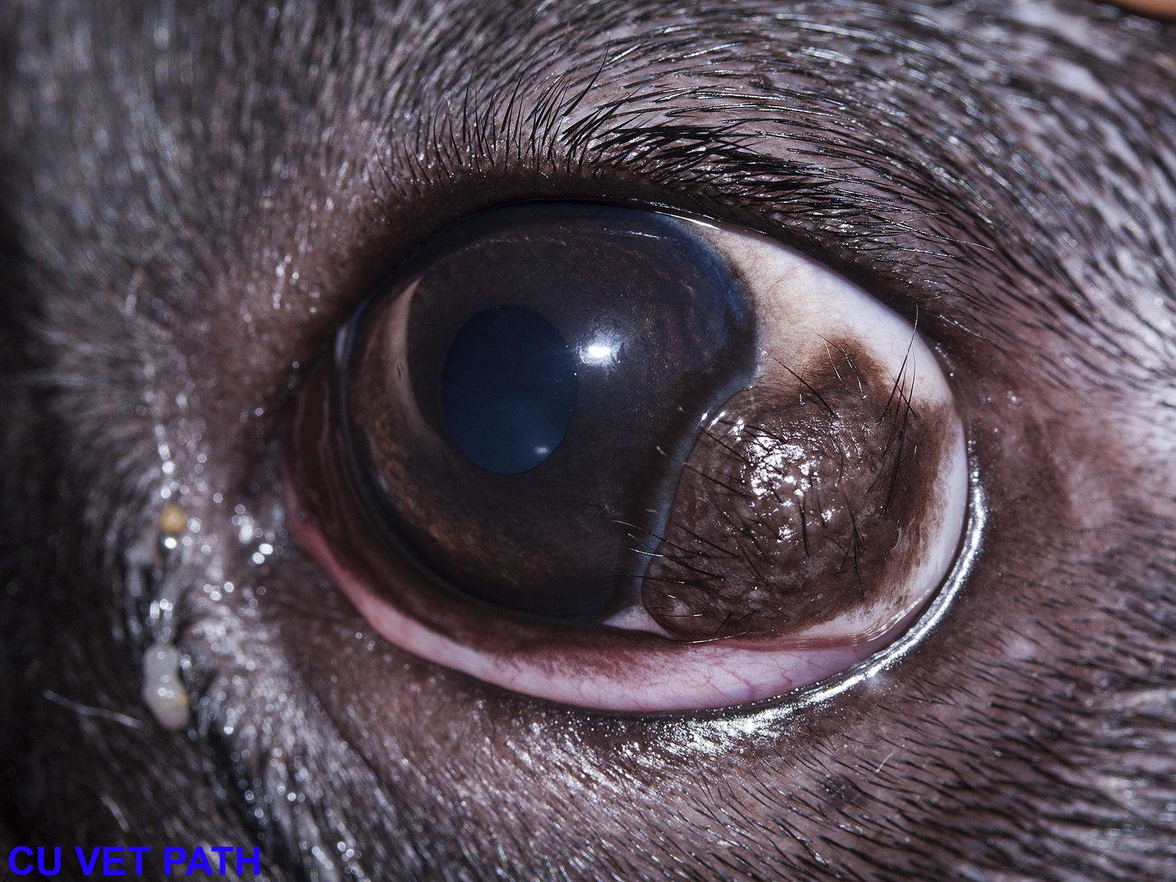
Dermoids are a form of choristoma: the formation of a normal structure in an abnormal place. In the case of a conjunctival (or corneal) dermoid, there is a nodule of skin present, which may be haired (Figure 2.1). These lesions are dramatic and odd, but are usually corrected through a keratectomy.

Figure 2.1: A dermoid present at the limbus (Image F33854 from Noah’s Arkive)
2.3 Meibomian gland adenoma
These are common neoplasms that occur at the eyelid margin, the only place in the body in which Meibomian glands are found. They make up almost 70 % of canine eyelid neoplasms, and are therefore the most common eyelid neoplasm of the dog. They are the eyelid equivalent to a cutaneous sebaceous adenoma, and are similarly benign and cured with complete resection. Grossly, they are usually well circumscribed and multilobular. Leakage of gland contents frequently creates a local granulomatous reaction that accompanies the neoplasm, known as a chalazion.
2.4 Conjunctival squamous cell carcinoma
Squamous cell carcinomas (SCC) of the conjunctiva or eyelid occur in all species, but are most common and most important in the horse and cow. In fact, SCC and Infectious bovine keratoconjunctivitis are the two most important ocular conditions of cattle. In both horses and cattle, UV light is thought to play a role in the pathogenesis, and those breeds with poorly pigmented skin are at increased risk. These neoplasms are infiltrative and have metastatic potential, though the true metastatic risk is poorly documented, and likely occurs late in disease. There is however a strong possibility that an animal may develop multiple conjunctival SCCs, and whether these separate masses are related is uncertain.
Grossly, conjunctival SCCs are typically raised, tan, and nodular. They are frequently ulcerated, with superifical crusts and hemorrhage. In cattle, they most frequently arise on the bulbar conjunctiva at the limbus, whereas the third eyelid is the most frequently affected in horses. In cats, SCC tends to arise from the skin of the eyelid. For unknown reasons, dogs seem relatively spared from developing SCCs.
2.5 Melanocytic tumours
Melanomas are frequent tumours of the eyelids and conjunctiva, and, in dogs, prognosis mostly depends on location. Melanocytic tumours arising at the eyelid margin are benign (like a dermal melanocytoma). Those that arise from the conjunctiva are more invasive and prone to recurrence (somewhat like oral melanomas). Those that arise from the limbus are benign.
2.6 Entropion and ectropion
Entropion refers to the inward folding of the eyelid, and is most common on the upper eyelid. Ectropion refers to an eyelid that turns slightly outward, and is more common in the lower eyelid. These are primarily clinical conditions and the underlying pathology is unremarkable. Both are most commonly seen in dogs, and there are strong breed predilections. Due to mechanical abraision, entropion may result in physical damage to the cornea. The condition is seen routinely and is easily fixed with minor surgery.